

For decades, the global response to Ebola outbreaks prioritized containment, viewing infected individuals primarily as biohazards rather than critically ill patients requiring active resuscitation (Fischer et al., 2019). Case fatality rates (CFRs) routinely hovered around 50% in resource-limited settings, largely driven by severe dehydration, electrolyte imbalances, and uncompensated hypovolemic shock (Jacob et al., 2020).
Conversely, the clinical management of severe Dengue has evolved into a highly systemized, protocol-driven discipline. Clinicians managing Dengue do not wait for cardiovascular collapse; they operate under a posture of expectant management, tracking subtle physiological indicators to guide volume-for-volume titration of fluids during a hyperacute critical phase.
While the exact viral triggers differ, the downstream physiological crises of severe Dengue and advanced EVD converge on endothelial dysfunction, massive fluid compartment shifts, consumption coagulopathy, and ischemic organ damage (Chavez et al., 2023; Wan et al., 2016).
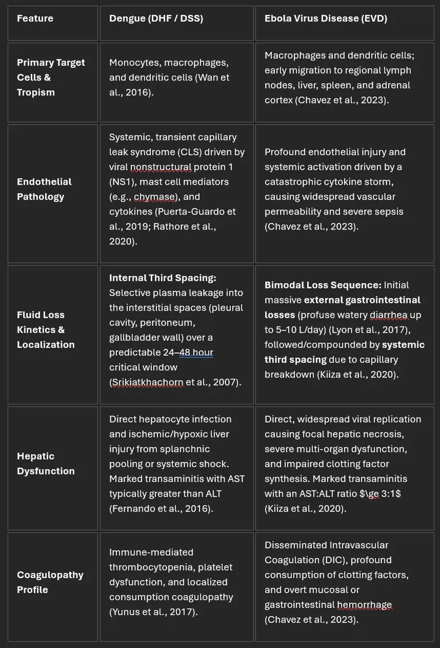
Translating Dengue Principles to Ebola Operational Protocols
1. Expectant Bedside Case Management & Tailored Observation Charts
The foundation of Dengue survival is a strict, structured tracking chart that records dynamic trends in volume status. In EVD, implementing a dedicated, high-visibility observation chart within the hot zone is vital.
Care teams must anticipate, rather than react to, hypovolemia by systematically tracking fluid input and output (intake of oral rehydration solution [ORS] vs. exact stool and vomit volumes) (Lyon et al., 2017). Vital signs must be trended to detect early, uncompensated hypovolemic shock (e.g., narrowing pulse pressure, postural heart rate spikes, or subtle changes in mental status) before blood pressure falls (Lamontagne et al., 2018).
2. Bedside Point-of-Care Ultrasound (POCUS) to Titrate Fluid Volumes
In Dengue, bedside ultrasonography is a crucial tool to detect early plasma leakage by visualizing subserosal fluid accumulation, such as gallbladder wall thickening, ascites, and pleural effusions (Srikiatkhachorn et al., 2007; Thulkar et al., 2000).
Tracking Internal Fluid Shifts: As EVD progress from pure gastrointestinal losses into secondary systemic vascular leak, POCUS can be rapidly deployed to identify internal third spacing. Volume Titration: Because advanced EVD patients are highly vulnerable to endothelial breakdown, aggressive, unmonitored fluid boluses risk inducing catastrophic pulmonary edema. Bedside POCUS allows clinicians to assess fluid responsiveness (via inferior vena cava respiratory variability) and monitor for early signs of fluid overload (such as standard lung interstitial fluid lines) (Kiiza et al., 2020).
3. Bedside Packed Cell Volume (PCV) to Decipher Hydration and Bleeding
Dengue management relies heavily on tracking serial hematocrit/PCV levels to balance plasma contraction (hemoconcentration) against hidden, internal bleeding (sudden drops in PCV) (Kularatne et al., 2021).
Quantifying Dehydration: In the hyperacute "wet" diarrheal phase of Ebola, point-of-care PCV testing provides an immediate estimate of hemoconcentration. High hematocrit values reflect severe intravascular volume depletion, indicating the need for immediate, volume-matched balanced crystalloid resuscitation (Lyon et al., 2017). Detecting Occult Blood Loss: Although EVD patients experience highly visible GI losses, internal or tissue-level bleeding can cause sudden cardiovascular collapse. Serial point-of-care PCV checks help clinicians differentiate between persistent hypovolemic shock (characterized by stable or rising, high PCV) and severe occult hemorrhage or DIC (indicated by a sudden drop in PCV relative to fluid intake), signaling the immediate necessity for blood component therapy instead of clear fluids (Lyon et al., 2017).
4. Utilizing Liver Function Tests (LFTs) for Metabolic Prognostication
In severe Dengue, the liver acts as a critical sentinel organ. Massive transaminase elevations (AST/ALT) serve as a proxy for hepatic ischemia and systemic hypoperfusion, accurately predicting multi-organ failure and death ( Devarbhavi et al., 2019).
Predicting Systemic Hypoxia: Point-of-care biochemistry panels in EVD should include serial liver enzyme tracking. An AST elevation disproportionate to ALT is a known prognostic marker of severe tissue hypoxia and viral necrosis in hemorrhagic fevers. Guiding Metabolic Interventions: Tracking these biochemical trends allows clinicians to anticipate metabolic crises, such as profound hypoglycemia or severe lactic acidosis, and initiate early corrections (e.g., hypertonic dextrose infusions) before irreversible multivisceral end-of-life failure occurs.
The most important clinical take-away
Clinical management of viral haemorrhagic fevers depends on the disciplined application of physiological principles at the bedside.
The task is simple in concept, but complex in execution: replace volume losses, correct electrolyte derangements, restore oxygen-carrying capacity when needed, and preserve vital organ perfusion long enough for the body — and available therapies — to fight the virus.
In this process, distinguishing fluid loss from blood loss is not just a technical skill; it is one of the critical arts of saving lives.
Dengue has taught us not to wait for collapse. Ebola supportive care needs the same bedside discipline: measure every loss, replace deliberately, reassess physiology continuously, and recognise the difference between dehydration and bleeding before organ failure becomes irreversible.
You finished · 3 min read
Originally published on LinkedIn by Dr Thisara Perera and Dr Yudara Kularathne MD, FAMS(EM).